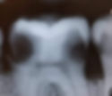
What a Cavity On a Molar Looks like
- David Chen, DDS
- Sep 20, 2022
- 7 min read
Cavities on molars look just like decay on any other teeth. The only difference is that this cavity is located on a molar tooth. Therefore the main differentiating factor is simply the location of the tooth decay. Aside from that, everything else looks practically the same.
Here are some general guidelines for what tooth decay looks like:
Shade of brown to black in color.
May have a cavitation (hole in the tooth).
Vary in size from small to large.
Can appear on any surface of the tooth - top, side, in between.
Texture may be hard or it could be soft.
Words are great but visual aid goes much further when it comes to identifying oral health problems such as decayed teeth. I'm sure you're very curious as to what cavities on molars look like. We're going to show you every variation of them.
Pictures of decay on molar teeth
Our molars do the bulk of the chewing, grinding, and mashing up of the food. They're very important teeth and since they're so crucial to eating, its no surprise that they can end up with cavities. They're the ones in contact with all that sugar that you're eating!
Here are pictures of every variation of cavities that you can possibly find on molars:
Cavity on the side of a molar
Decay can develop on the side of your molar, either on the cheek side or the tongue side. Although it is more common on the cheek side. If it does form, you'll notice a brown spot or dot that is at the groove in the middle of the tooth. It can form outside of that grove but due to its shape it just tends to be more prone to decay.

Treatment for these is usually just a simple cavity filling. If its shallow enough you may not even need to be numb for it. Although if you do feel something, you can always let your dentist know and they'll give you some Lidocaine to numb it.
Cavity on the chewing surface of a molar
The top of the tooth is where the mastication or chewing is done. That makes it easy for food to get stuck in the grooves and end up forming a cavity. It will look like a black dot in one of the grooves in the tooth.

If you catch these early, you can just do a tooth filling on it. However if you allow it to grow, it will get much bigger than what you see above. Instead of a small dot it can encompass a much greater surface area.
Here is an example of how big it can grow. As you can see that small brown dot has become a sizable big brown circle.

Cavity that is big enough to be into the nerve
Decay that is left untreated will eventually reach the nerve of the tooth. Once it does it will start causing you pain. The reason why most people tend to ignore small cavities is because they're often painless. However, once they get close to the nerve it will start causing you pain and that is when people are usually unable to ignore them.
For a cavity to reach the nerve, they usually have to grow to about this size. The entire tooth may be cavitated and they'll look like a dark brown to black color.

Once the decay has reached this size, a composite filling will be insufficient. This tooth will at least need a root canal and a crown. However, there are times where the decay is just so extensive that by the time it is completely cleaned out there will be not enough tooth structure left to put a crown on.
If there isn't enough tooth to put a crown on, you may need the entire tooth extracted instead. For this reason alone, you should never leave small cavities untreated. You should deal with them as soon as possible so that you can save yourself time and money.
Cavity underneath an existing molar filling
Just because you had that cavity filled, it does not make it exempt from future decay. Yes, you can get another cavity underneath a cavity filling! Whenever that happens we don't call it regular tooth decay but it is instead referred to as "recurrent decay". That means that it is not the first time that you've had it happen to that tooth.
Here is a picture of what recurrent decay looks like:

What it looks like is an old cavity filling that has discoloration underneath of it. It will look brown or black underneath the filling material. Whenever you see this, you should be immediately suspicious of decay that has snuck in under the filling material.
This can be confirmed by the fact that in the filling you can also see a crack running through it. The bacteria most likely got in through the crack and edges of the restoration and caused a cavity to form.
The treatment for this would be to replace the filling with a brand one one. Your dentist will most likely need to clean out the rotten part of the tooth that is underneath of it as well prior to putting a new restoration in.
Cavity from a broken filling on a molar
Old fillings can eventually start breaking down. Once it does, cracks will start forming and it is through these cracks that bacteria and decay can get in. Once it does, it will start causing a change in color in the tooth structure. It'll usually start looking like a brown or black color.

Due to this reason alone, once you notice your filling starting to break down, you should try to replace it as soon as possible. You want to keep it as a small problem and not let it progress to something larger like a root canal.
Decay that is in between the molar surfaces
If you're not too thrilled with flossing, the molars can end up with decay in between them. The unfortunate part of this type of cavity is that you're unable to detect them with the naked eye. The only way to know if you have these is if you take annual x-rays.

What it'll look like on the x-ray is black circles in between the teeth. Imagine where you would floss through the molar and that is where you'll see the dark circles. The larger the circles, the bigger the decay.
Pictures of decay on wisdom teeth
It is quite common for wisdom teeth (third molars) to get cavities. In fact they're more prone to getting decay because of how far back they're located which adds to the challenge of keeping them clean. To make matters worse, a lot of wisdom teeth grow in impacted so that only adds an additional barrier to oral hygiene.
Here are pictures of wisdom teeth with cavities on them that were taken at our long island city dental office.
Decayed chewing surface of a third molar
The surface where you chew your food can become rotten. Once it does the color will change to a brown-black shade. It is a sign that you're not able to keep this tooth clean since it is so far back in your mouth. Perhaps you're not brushing far back enough?

These teeth could potentially be treated with a cavity filling. Although most people simply opt to just have them extracted since the primary problem is being able to keep them clean in the first place.
Cavity on the side of a third molar
This picture has a big cavity on the cheek side of the wisdom tooth. The decay is so large that it actually formed a cavitation in the tooth, which is an actual hole. That hole is brown in color and that tells you it is definitely rotten. The rest of the tooth isn't that color.

This condition is a result of not getting your toothbrush far back enough to clean them. Most people are unable to see their wisdom teeth so they're never quite sure if they're actually cleaning them. Our advice is to push the brush a little bit further back if you think you've gone far back enough!
This tooth can potentially be restored with a filling but due to the size, we would recommend to have it removed instead. Due to how far back the tooth is, it makes it hard for your dentist to work on it. It is simply easier to have it extracted.
Cavitated wisdom tooth
Untreated tooth decay will eventually lead to the formation of a large cavity. This wisdom tooth was such a case, where a large hole formed. The decay weakened the structural integrity so much that part of the tooth just came off thus leaving a hole.

If your wisdom tooth is this broken down like the photo above, it is simply impossible to save it and restore it with a tooth filling. This tooth will definitely need to be extracted so you should prepare yourself!
Alternatively, you can try doing a root canal and crown on it... but the success rate for that on third molars is questionable. Both of those procedures would cost significantly more than a wisdom tooth extraction. You may be wasting your time and money if you do that. We would have to recommend against doing so.
Learn more: Cost of a root canal and crown.
First of all, you don't really chew with your third molars so we don't believe it's the best use of your money and time.
Takeaway
Decayed molars look very similar to other teeth that have cavities. The only difference is that this type of cavity is simply located on your molars. The reason is because the stages of tooth decay remain the same regardless of what teeth they affect.
Nonetheless, it is still helpful to know what they look like, which was the purpose of this article. To show you what every form and variation of molar decay can look like. Hopefully we've fulfilled your desire and if you need a dental filling in long island city, we can certainly help you with that.
Learn more: If you enjoyed reading about this, you should also check out our full article on what cavities look like.
